
Blog Post
What is Digital Pathology
Proscia Staff
05 Jan 2026

Digital Pathology Overview
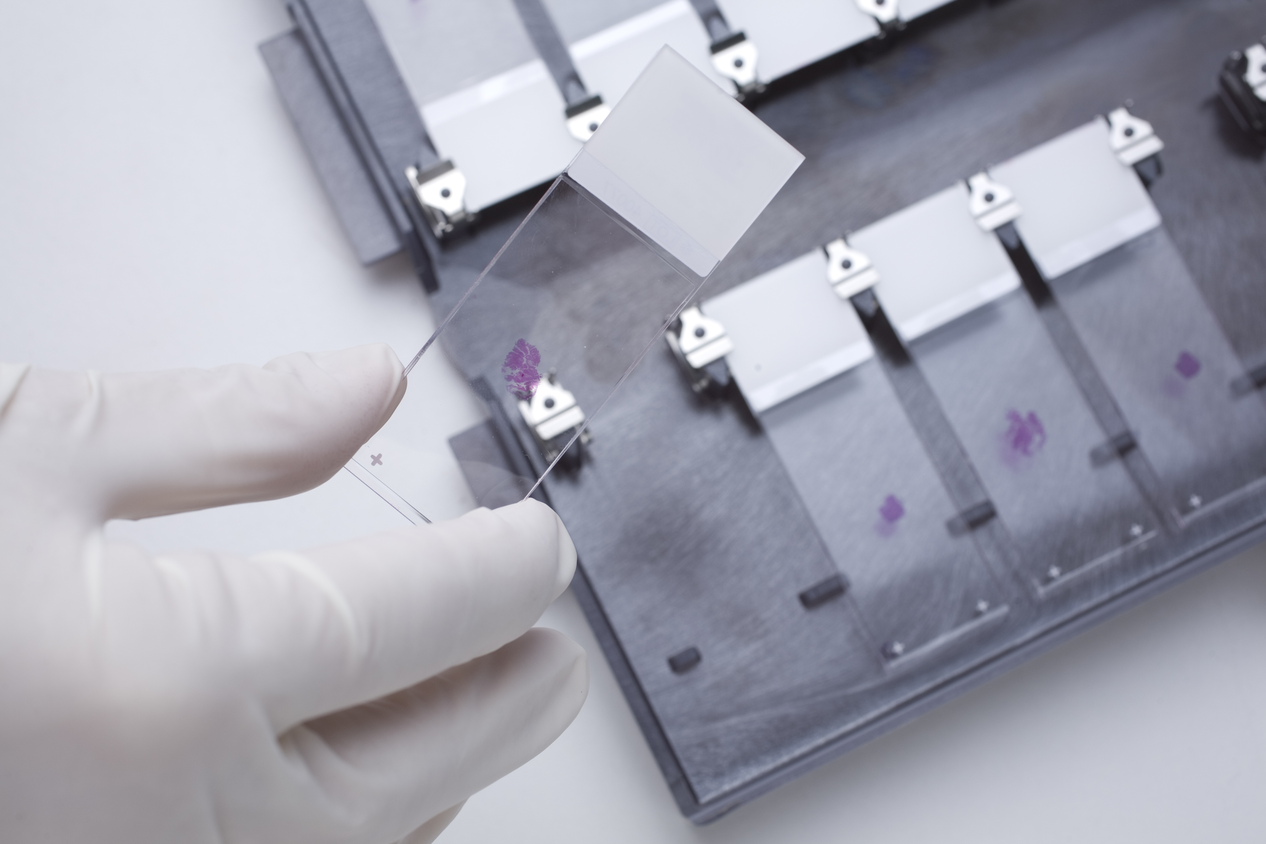
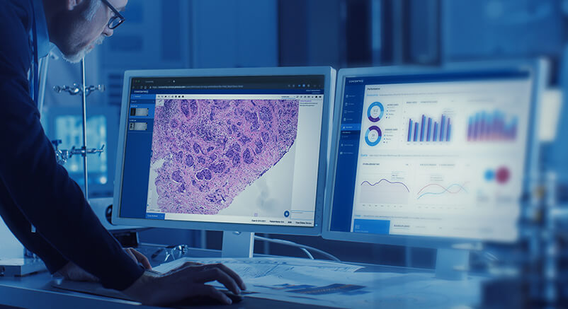
Digital pathology is the process of digitizing glass slides using a whole slide image scanner and then analyzing the digital images using an image viewer, typically on a computer monitor or mobile device. An image viewer works similarly to the traditional standard light microscope enabling pathologists to move slides around in the same way. Although the basic viewing functionality has not changed drastically, digital pathology has brought about amazing advancements for pathology lab efficiency, workflow, and revenue enhancements.
The evolution of digital pathology, or whole slide imaging (WSI), has taken almost 20 years. It has progressed from attaching a camera to the lens of a microscope, to the development of the first scanners, to what we have today: technology that is quickly becoming indispensable in the anatomic laboratory… The first scanners were clunky instruments taking up large footprints – with long processing time (up to 6 minutes) expensive storage, and limited use cases. Scanners today can scan slides in as little as 30 seconds, be configured at multiple magnifications, and handle up to 1,000 slides at a time. Storage, now available in the cloud, is affordable, secure, and accessible. Computational applications incorporating artificial intelligence (AI) offer a myriad of ways to analyze and present images to the pathologist.
The evolution of digital pathology is quite similar to that of the mobile phone. Early versions were unnecessarily big, eccentric, and limited by coverage, cost, and need. Now, the mobile phone is as ubiquitous as the watch and used in ways no one could have imagined in the early 1980s. With the evolution of whole-slide image scanners, digital pathology as a practice has taken off, much like mobile phone usage has skyrocketed with more powerful mobile devices.
How is digital pathology used today?
There are three main ways in which digital pathology is used today. First, research institutions, such as pharmaceutical companies, CROs, and academic medical centers, use digital pathology in robust study design, data collection, and database management for the millions of specimen that they currently manage and seek to leverage.
Second, clinical labs use digital pathology on select cases for remote consultations, education, or quantitative analysis.
The third and fastest-growing use case centers on clinical labs moving to an all-digital workflow. These labs use computatioanlly-enabled digital pathology powered by AI to help assign cases to pathologists, manage the workflow into worklists, offer quantitative image analysis for specific case types, integrate information into their existing Lab Information System (LIS), and expand access to cases beyond the brick-and-mortar walls of the lab itself. With digital pathology, diagnosis no longer needs to be delayed by the physical shipment of tissue samples or the wait times that result when pathologists are out of the office.
How does computational pathology enhance the value of whole slide imaging?
AI applications can “read” a whole slide image and apply specialized algorithms that can perform many useful clinical tasks to augment the role of the pathologist. It is well known that there is fundamental prognostic data embedded in pathology images. Software applications can now quantify aspects of tissue often invisible to humans even under a microscope to predict the likely diagnosis, the aggressiveness of tumors, and ultimately, patient outcomes. These AI applications, under the umbrella of computational pathology, are redefining why labs are quickly adopting digital pathology. The same applications can be used to reduce malpractice by offering the equivalent of a second opinion, detecting patterns that the human eye can’t see, or alerting pathologists of any discrepancies.
There are also other use cases, such as sorting and workload balancing, that computational applications can help to perform in the background to make the laboratory workflow more efficient and make the best use of pathologists’ time. For example, an AI application can auto-categorize tissue samples by disease state and then route them to specific pathologists for review. A more senior pathologist could assess the straightforward cases first, saving the more complex biopsies for later in the day.
Conversely, a pathologist could assign straightforward cases to his less experienced colleagues, tackling more complex cases himself right out of the gate. Finally, there are now avenues for increasing revenue using digital pathology as a means to attract clients that want to read their own cases but don’t have a laboratory for preparing slides as seen in this example.
Digital Pathology and AI
What do people mean by digital pathology?
Digital pathology is the practice of converting glass slides into high-resolution digital images and using software to view, manage, share, and analyze those slides.
Instead of relying solely on microscopes and physical slides, teams can work with digital slides on a screen—enabling easier access, faster collaboration, and a foundation for computational analysis. Digital pathology is not just an IT upgrade. It enables workflows and insight that are difficult to achieve with glass alone.
What is a whole slide image, and why does it matter?
A whole slide image (WSI) is a high-resolution digital scan of an entire glass pathology slide. WSIs typically reach 1–3 gigapixels per slide, capturing tissue at multiple magnifications and generating 1–4 GB of data per image, depending on compression and modality.1,2
Unlike a single static image, a WSI captures the full slide at multiple magnifications, allowing users to pan and zoom much like navigating a map. WSIs are the building blocks of digital pathology—once slides are digitized, teams can review them remotely, share them quickly, and apply computational tools, including AI, to analyze them.
How is digital pathology helping teams manage growing workloads and limited staffing?
Digital pathology helps teams handle increasing case volumes and staffing constraints by improving efficiency, enabling remote collaboration, and supporting more scalable workflows across sites and teams.
In practice, this includes:
- Remote and distributed work, allowing pathologists to collaborate across sites and cover gaps more flexibly
- Workflow efficiency, reducing time spent on manual slide handling and case retrieval
- Scalability, so rising volumes don’t require linear increases in staffing
- AI-assisted support, which can help prioritize work and reduce variability for specific tasks
Digital pathology doesn’t eliminate staffing challenges, but it helps teams work more sustainably under growing operational pressure.
Where does digital pathology create the most value?
Digital pathology creates value in practical, measurable ways:
- Efficiency: faster case access, fewer manual steps, streamlined workflows (often measured in minutes saved per case, which compounds at enterprise scale)
- Collaboration: simpler consultation and subspecialty review across locations
- Consistency: more standardized workflows and easier quality programs
- Scalability: support for growing case volume and distributed teams
- Insight: the ability to extract quantitative information from tissue at scale
These benefits appear across clinical practice, research, and drug development—often with different starting points and success metrics.
How are pathology teams actually using digital pathology today?
Clinically, digital pathology is often adopted to improve workflow and collaboration first, then expanded to support quality, operational visibility, and scale.
Common clinical uses include:
- Primary diagnostic review (where permitted or cleared)
- Subspecialty consultation and second opinions
- Tumor boards and multidisciplinary review
- Multi-site standardization
In enterprise deployments, the most successful programs prioritize pathologist workflow and adoption early—because if pathologists don’t want to use the system, nothing else matters.
Notably, industry sources report that roughly one-third of clinical laboratories have begun or are planning whole-slide imaging.3
How are life sciences teams using digital pathology?
In the life sciences, digital pathology is less about diagnosis and more about turning tissue into scalable data.
Pharma and biotech teams use digital pathology across discovery, translational research, and clinical development to analyze tissue more consistently and quantitatively. Common use cases include:
- Biomarker discovery and validation
- Spatial biology and tissue microenvironment analysis
- Toxicologic pathology in preclinical safety studies
- Companion diagnostics development
- Clinical trial enablement and standardization
Where does AI actually fit into digital pathology workflows?
AI builds on digital pathology—it doesn’t exist without digitized slides.
Once tissue is captured as WSIs, AI can assist with well-defined tasks such as detection, quantification, and spatial analysis. The most practical way to think about AI is as decision support: it helps pathologists and scientists work more efficiently and consistently, while keeping human judgment at the center.
Because AI is evolving rapidly, platforms should support flexibility—multiple AI applications, including third-party tools, integrated directly into workflows rather than treated as separate tools.
Is AI being used to replace pathologists?
No. AI in pathology is designed to support pathologists, not replace them.
Pathology relies on clinical context, judgment, and experience.While there are a handful of digital pathology and pathology-AI products that have received regulatory clearance, While AI is best suited to narrow, structured tasks that reduce variability and workload, allowing pathologists to focus more time on interpretation and complex decision-making.
How reliable is AI in real-world pathology settings?
AI accuracy depends on context.
Performance is shaped by training data diversity, validation for a specific use case, and verification in the environment where it’s deployed. Accuracy is not a single universal number—it must be evaluated based on intended use and monitored over time as workflows, scanners, and populations change.
When teams adopt digital pathology, what should come first—software or scanners?
In most cases, organizations should choose the software platform first, then select scanner hardware that fits their workflows and scale.
The software platform defines how work gets done, how data flows, and which integrations and AI capabilities are possible. Scanner hardware is essential, but interchangeable. A hardware-first approach often leads to vendor lock-in and limits flexibility over time.
A software-first approach helps organizations:
- Keep scanners interchangeable across sites and use cases
- Adopt multiple AI applications as they become available
- Avoid proprietary constraints and costly migrations
What should teams prioritize when evaluating a digital pathology platform?
Look for a platform that is built for pathologists, open to scanners and AI, and ready for enterprise integration and scale over time—not just a basic image management system.
Key criteria to prioritize:
- Pathologist-first experience: fast, intuitive, microscope-like navigation
- Openness and interoperability: scanner-agnostic support and standards alignment
- AI flexibility: support for multiple concurrent AI applications, including third-party tools
- Enterprise integration: proven bi-directional LIS integration
- Future readiness: support for research, real-world data, and life sciences use cases without separate platforms
Do most organizations rely on a single vendor for all digital pathology needs?
Usually, no—and that’s often a good thing.
Digital pathology is an ecosystem that includes scanners, software platforms, integrations, AI applications, storage, and research workflows. Some vendors attempt to bundle everything, but tightly bundled ecosystems can limit choice, restrict AI adoption, and make it harder to evolve over time.
A more resilient, long-term approach is to select an open, enterprise software platform that can integrate with:
- Multiple scanner manufacturers and formats
- Multiple AI applications, including third-party tools
- Enterprise systems such as the LIS and EHR
This allows organizations to choose best-fit components while keeping the platform as a stable foundation that can evolve as needs change.
AI Biomarker Detection in Cancer: Common Questions, Answered
What are biomarkers in cancer?
Biomarkers are measurable characteristics that provide information about a biological process, disease state, or response to therapy.
In cancer, biomarkers can indicate the presence of disease, help classify tumor subtypes, predict prognosis, or guide treatment decisions. They may be molecular (such as genetic mutations or protein expression), cellular, or tissue-based—and they are increasingly central to precision oncology.
Pathology has long played a critical role in biomarker assessment, particularly through tissue evaluation, where the structure, composition, and spatial organization of cells provide essential diagnostic and prognostic insight.
What are tissue-based biomarkers?
Tissue-based biomarkers are features observed directly within tissue samples, typically assessed using histopathology, immunohistochemistry (IHC), or in situ hybridization.
Examples include:
- Protein expression levels (e.g., receptor status)
- Cell counts or proportions
- Morphologic patterns
- Spatial relationships between cells and structures
- Features of the tumor microenvironment
Unlike blood-based biomarkers, tissue-based biomarkers preserve spatial and architectural context—information that is often critical for interpretation and clinical relevance.
How are biomarkers traditionally identified in pathology?
Traditionally, tissue biomarkers are identified through manual review by pathologists, often supported by special stains or IHC.
This process relies heavily on expert visual assessment and semi-quantitative scoring. While highly valuable, it can be:
- Time-intensive for high-volume studies
- Subject to inter- and intra-observer variability
- Difficult to scale consistently across large datasets or multi-site trials
As studies grow larger and more complex, these limitations become more pronounced—particularly in research and clinical trial settings where reproducibility and throughput are essential.
What is AI-based biomarker detection?
AI-based biomarker detection uses computational models to identify, quantify, and analyze features within digitized tissue slides.
Once slides are scanned into whole slide images (WSIs), AI can be applied to perform tasks such as:
- Detecting specific cell types or structures
- Quantifying biomarker expression across entire slides
- Measuring spatial relationships within tissue
- Identifying patterns that may be subtle or difficult to assess visually
Rather than replacing pathologists, AI is used to support more consistent, scalable, and quantitative analysis—especially for well-defined tasks.
Why does digital pathology matter for AI biomarkers?
AI cannot be applied directly to glass slides. Digital pathology is the foundation.
By converting slides into high-resolution WSIs, digital pathology enables:
- Computational access to tissue data
- Standardized image inputs for AI models
- Scalable analysis across thousands of slides
- Integration of AI results directly into review workflows
Without digital pathology, AI-based biomarker detection is not possible at scale. The quality, consistency, and accessibility of digitized slides directly affect AI performance and reliability.
What types of biomarkers are well-suited for AI detection?
AI performs best on structured, repeatable tasks with clear definitions. In pathology, this often includes:
- Cell detection and classification
- Quantification of staining intensity or proportion
- Counting positive vs. negative cells
- Measuring spatial features and distributions
- Segmenting tissue regions
These tasks are particularly valuable in studies where consistency and throughput are critical, such as biomarker discovery, translational research, and clinical trials.
More complex interpretive judgments remain the responsibility of pathologists, with AI serving as an assistive tool.
How are AI biomarkers used in cancer research and drug development?
In the life sciences, AI-based biomarkers help turn tissue into scalable data.
Common applications include:
- Biomarker discovery and validation
- Patient stratification for clinical trials
- Endpoint assessment and response evaluation
- Spatial biology and tumor microenvironment analysis
- Companion diagnostics development
By applying consistent algorithms across large cohorts, teams can reduce variability, improve statistical power, and generate insights that are difficult to achieve with manual review alone.
Are AI biomarkers used clinically today?
Some AI-based pathology tools have received regulatory clearance for specific clinical use cases, including certain biomarker assessments.
However, clinical adoption varies by region, indication, and regulatory environment. In practice, AI biomarkers are most commonly introduced as decision support—helping pathologists work more efficiently and consistently while maintaining human oversight.
Clinical deployment requires careful validation, workflow integration, and ongoing performance monitoring.
How reliable are AI-based biomarkers?
AI reliability depends on context.
Key factors include:
- Diversity and quality of training data
- Validation for the intended use case
- Alignment with local scanners, stains, and workflows
- Ongoing monitoring after deployment
There is no single accuracy number that applies universally. Performance must be evaluated in the environment where the AI is used and revisited as conditions change.
What role do pathologists play in AI biomarker workflows?
Pathologists remain central.
They define the clinical question, validate results, interpret findings in context, and make final decisions. AI helps by reducing manual burden, improving consistency, and surfacing quantitative data—but it does not replace professional judgment.
Successful AI biomarker programs are those that are designed around pathologist workflows, not layered on top of them as disconnected tools.
What should teams look for in a platform supporting AI biomarkers?
Teams should prioritize a digital pathology platform that supports flexibility, integration, and scale over time.
Key considerations include:
- High-performance whole slide image viewing
- Support for multiple AI applications, including third-party tools
- Integration of AI results directly into review workflows
- Interoperability with scanners and enterprise systems
- Ability to support both clinical and life sciences use cases
Because biomarker strategies evolve, platforms should be built to adapt—not lock teams into a single algorithm or vendor.
Where is AI biomarker detection headed next?
AI biomarker detection is moving toward greater scale, richer spatial analysis, and tighter integration across research and clinical workflows.
As digital pathology adoption grows, tissue data will increasingly serve as a bridge—connecting discovery, development, and clinical care. AI will play a growing role in unlocking that data, while pathologists remain essential to ensuring its responsible and meaningful use.
Where is digital pathology headed?
As AI-enabled digital pathology continues to advance, it will likely follow most technology curves; the costs will continue to come down and the variety of applications will increase. The declining number of pathologists combined with the rise in biopsy volume is necessitating the need for more efficient labs. Additionally, crossover with other AI applications, like those in radiology and MRI, will provide opportunities to use digital pathology tools to as part of integrated diagnostics and prognostic decision making.
Some labs have already set up their digital pathology system to auto-load select images from their radiology PACS (Picture Archiving Communication System) upon case signout, allowing treating physicians to view and navigate the very slide from which patient diagnosis is based. At the same time, radiologists can see radiographs along with MRI and CT scans and the corresponding histology slides in one view.
With so many examples of labs doing great things by adopting digital pathology, suddenly the hurdles of increasing server storage and rearranging infrastructure seem less daunting than before. More and more labs will be dusting off their scanners and reengaging in the conversation to evolve to digital pathology. As the adoption of digital and computational pathology grows, labs will experience more dramatic increases in workflow efficiency and enjoy a new era of collaboration. The ultimate outcome? Faster, more accurate diagnoses for patients, and a broader volume of information at their treating physicians’ fingertips.
At Proscia, we believe that pathology deserves great technology.
References
Ashman K, Zhuge H, Shanley E, et al. Whole slide image data utilization informed by digital diagnosis patterns. J Pathol Inform. 2022;13:100113. Published 2022 Jun 30. doi:10.1016/j.jpi.2022.100113
- Hancox J. Whole Slide Image Analysis in Real Time with MONAI and RAPIDS. NVIDIA Technical Blog. July 13, 2023. Updated November 20, 2023. Accessed January 9, 2026. https://developer.nvidia.com/blog/whole-slide-image-analysis-in-real-time-with-monai-and-rapids
- Wallask S. Slow Digital Pathology Adoption Continues, According to Labcorp Report. Today’s Clinical Lab. Published January 30, 2025. Accessed January 9, 2026. https://www.clinicallab.com/slow-digital-pathology-adoption-continues-according-to-labcorp-report-28167