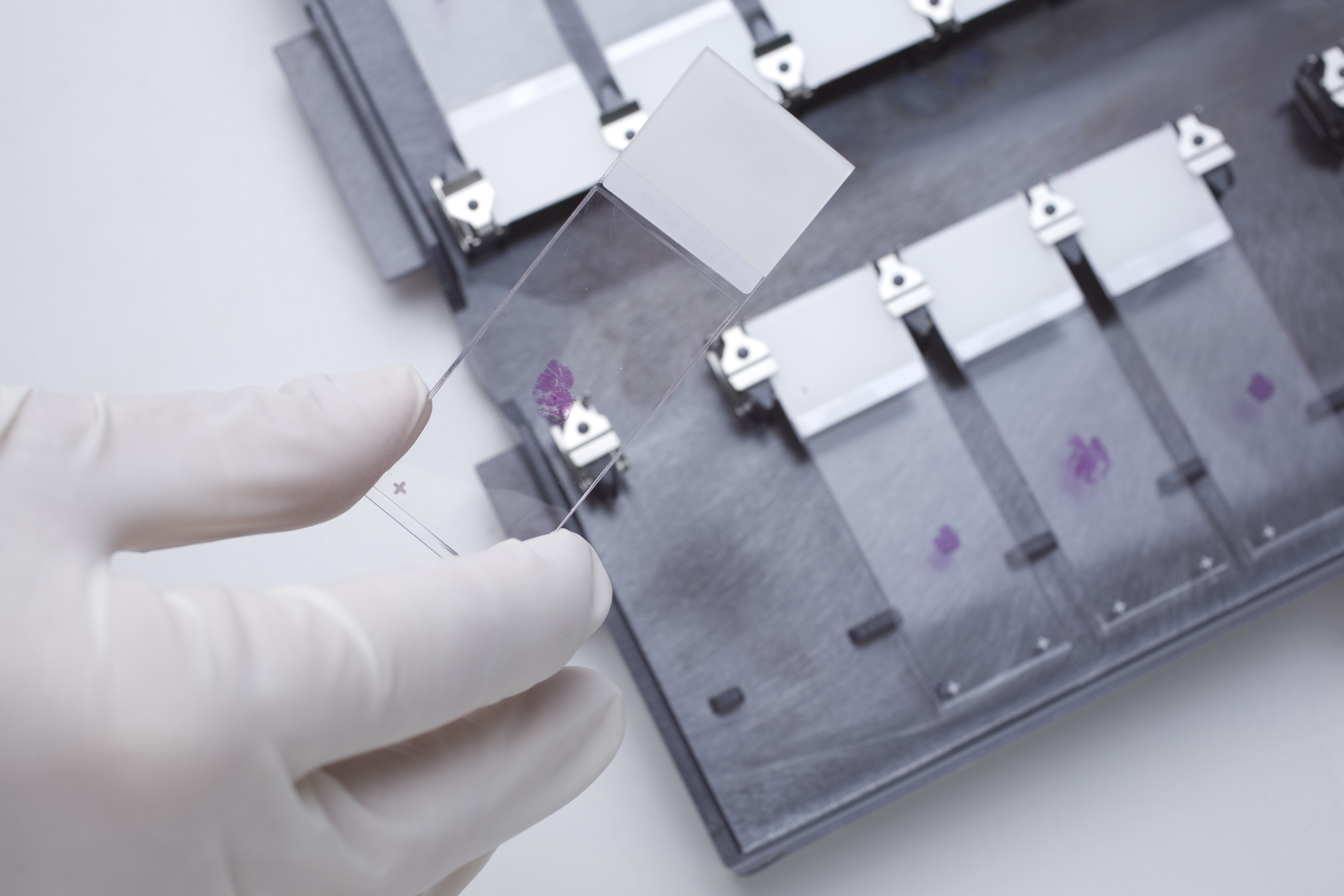
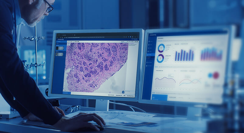
Digital pathology is the process of digitizing glass slides using a whole slide image scanner and then analyzing the digital images using an image viewer, typically on a computer monitor or mobile device. An image viewer works similarly to the traditional standard light microscope enabling pathologists to move slides around in the same way. Although the basic viewing functionality has not changed drastically, digital pathology has brought about amazing advancements for pathology lab efficiency, workflow, and revenue enhancements.
The evolution of digital pathology, or whole slide imaging (WSI), has taken almost 20 years. It has progressed from attaching a camera to the lens of a microscope, to the development of the first scanners, to what we have today: technology that is quickly becoming indispensable in the anatomic laboratory… The first scanners were clunky instruments taking up large footprints – with long processing time (up to 6 minutes) expensive storage, and limited use cases. Scanners today can scan slides in as little as 30 seconds, be configured at multiple magnifications, and handle up to 1,000 slides at a time. Storage, now available in the cloud, is affordable, secure, and accessible. Computational applications incorporating artificial intelligence (AI) offer a myriad of ways to analyze and present images to the pathologist.
The evolution of digital pathology is quite similar to that of the mobile phone. Early versions were unnecessarily big, eccentric, and limited by coverage, cost, and need. Now, the mobile phone is as ubiquitous as the watch and used in ways no one could have imagined in the early 1980s. With the evolution of whole-slide image scanners, digital pathology as a practice has taken off, much like mobile phone usage has skyrocketed with more powerful mobile devices.
How is digital pathology used today?
There are three main ways in which digital pathology is used today. First, research institutions, such as pharmaceutical companies, CROs, and academic medical centers, use digital pathology in robust study design, data collection, and database management for the millions of specimen that they currently manage and seek to leverage. Learn about Solutions for Life Sciences organizations.
Second, clinical labs use digital pathology on select cases for remote consultations, education, or quantitative analysis.
The third and fastest-growing use case centers on clinical labs moving to an all-digital workflow. These labs use computatioanlly-enabled digital pathology powered by AI to help assign cases to pathologists, manage the workflow into worklists, offer quantitative image analysis for specific case types, integrate information into their existing Lab Information System (LIS), and expand access to cases beyond the brick-and-mortar walls of the lab itself. With digital pathology, diagnosis no longer needs to be delayed by the physical shipment of tissue samples or the wait times that result when pathologists are out of the office. Learn about Solutions for Diagnostic Labs.
How does computational pathology enhance the value of whole slide imaging?
AI applications can “read” a whole slide image and apply specialized algorithms that can perform many useful clinical tasks to augment the role of the pathologist. It is well known that there is fundamental prognostic data embedded in pathology images. Software applications can now quantify aspects of tissue often invisible to humans even under a microscope to predict the likely diagnosis, the aggressiveness of tumors, and ultimately, patient outcomes. These AI applications, under the umbrella of computational pathology, are redefining why labs are quickly adopting digital pathology. The same applications can be used to reduce malpractice by offering the equivalent of a second opinion, detecting patterns that the human eye can’t see, or alerting pathologists of any discrepancies.
There are also other use cases, such as sorting and workload balancing, that computational applications can help to perform in the background to make the laboratory workflow more efficient and make the best use of pathologists’ time. For example, an AI application can auto-categorize tissue samples by disease state and then route them to specific pathologists for review. A more senior pathologist could assess the straightforward cases first, saving the more complex biopsies for later in the day. Learn more about AI Applications.
Conversely, a pathologist could assign straightforward cases to his less experienced colleagues, tackling more complex cases himself right out of the gate. Finally, there are now avenues for increasing revenue using digital pathology as a means to attract clients that want to read their own cases but don’t have a laboratory for preparing slides as seen in this example.
Where is digital pathology headed?
As AI-enabled digital pathology continue to advance, it will likely follow most technology curves; the costs will continue to come down and the variety of applications will increase. The declining number of pathologists combined with the rise in biopsy volume is necessitating the need for more efficient labs. Additionally, crossover with other AI applications, like those in radiology and MRI, will provide opportunities to use digital pathology tools to as part of integrated diagnostics and prognostic decision making.
Some labs have already set up their digital pathology system to auto-load select images from their radiology PACS (Picture Archiving Communication System) upon case signout, allowing treating physicians to view and navigate the very slide from which patient diagnosis is based. At the same time, radiologists can see radiographs along with MRI and CT scans and the corresponding histology slides in one view.
With so many examples of labs doing great things by adopting digital pathology, suddenly the hurdles of increasing server storage and rearranging infrastructure seem less daunting than before. More and more labs will be dusting off their scanners and reengaging in the conversation to evolve to digital pathology. As the adoption of digital and computational pathology grows, labs will experience more dramatic increases in workflow efficiency and enjoy a new era of collaboration. The ultimate outcome? Faster, more accurate diagnoses for patients, and a broader volume of information at their treating physicians’ fingertips.
At Proscia, we believe that pathology deserves great technology. Learn more about us.


