The 2022 release of Concentriq for Research is in line with Proscia’s mission to develop robust technology to help our customers and prospects to accelerate pathology image based research to promote the incredible breakthroughs in research for diseases like cancer.
This newest launch of Concentriq for Research focuses on two major enhancements critical to driving cutting-edge high-throughput research: expanded fluorescence support, and the introduction of AI-enabled automated quality control. Fluorescent imaging is one of the most widely used techniques in life sciences research from cancer research, immunology, to neuroscience and more. The latest launch brings fluorescent imaging into the enterprise image management system trusted by 10 of the top 20 life sciences companies to power their research. Additionally, Concentriq for Research now supports automated quality control that leverages AI trained and tested on thousands of samples to identify commonly-occurring quality issues in every image of H&E stained slides, reducing dependency on a manual, tedious tasks and helping Concentriq users drive faster, high-quality research. Let’s dive a bit deeper into these and other features and enhancements now available on Concentriq.
Empowering fluorescence image based research with the latest version of Concentriq for Research
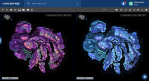
To support spatial biology research in the life sciences and provide tools to assist with the breakthrough innovations, Concentriq for Research now includes support for fluorescence in the same mature enterprise grade platform that has enabled image-based research for some of the largest global teams. Key fluorescence improvements now available in Concentriq include:
- An intuitive workflow for fluorescent image viewing: Users are now able to compare multiple fluorescent and brightfield images side-by-side in Concentriq’s multiview. Additionally, biomarker names – separable from channel names – are now visible alongside the images, giving researchers additional context about the images they’re analyzing. Other, general interface enhancements – including an easy-to-use intensity adjuster – deliver a simplified and efficient workflow for researchers who review fluorescent images regularly.
- Easy incorporation and access of fluorescence images and fields alongside brightfield and other imaging data: The latest launch allows users to leverage the power of Concentriq’s robust global search functionality on fluorescent images. The images are now searchable by biomarkers, allowing the organization to easily access their past and present data. This metadata is organized and managed alongside other pathology data, making it easier than ever to bring the power of fluorescent imaging to a broader body of R&D.
- Standardized fluorescent images and data management across the organization: Support for fluorescence in project templates and metadata field libraries encourages the collection of consistent data across studies. Concentriq for Research allows admin users to create and manage a library of metadata fields for the organization, specifying what the metadata field applies to (such as folder, slide, or annotation) and in what form the data is captured (such as dropdown, checkbox, date, number, and text). The platform also enables users to create metadata templates, which can be applied to studies of similar types repeatedly. By expanding this platform functionality to fluorescent images, researchers can better incorporate this imaging modality alongside other pathology data.

Check out the product tour hosted by Nathan Buchbinder and Edaeni Hamid to learn more about the latest features of Concentriq for Research 3.7.
Proscia Introduces AI-Powered Quality Control to Accelerate Data-Driven Drug Development
Life sciences organizations face unprecedented pressure to rapidly develop novel therapies, as the COVID-19 pandemic has highlighted the potential of the digital transformation in advancing breakthroughs that fight disease. While these organizations have been on the forefront of digital pathology adoption, generating millions of images to fuel a new wave of research, the images often contain quality issues that delay researchers from realizing their full value.
Automated quality control (QC) leverages AI that has been trained and tested on thousands of samples to identify commonly-occurring quality issues in every image of H&E stained slides. The AI-enabled workflow automation application detects the many of the most frequently-occurring quality artifacts that necessitate a re-prep or rescan, including those that result both from slide preparation and from the use of WSI scanners. It plugs seamlessly into the research workflow powered by Concentriq for Research. By reducing the need to manually review these images, it drives significant productivity and quality gains that may enable research teams to:
- Start studies faster: AI that automatically flags common quality artifacts, from air bubbles to blurriness, gives researchers faster access to high-quality data.
- Increase the reproducibility of results: Reliable research results depend on the quality of the data that informs them. The application’s highly accurate AI ensures consistent quality across even the largest data sets.
- Reduce technician burnout: The burden of manual quality control is intensifying as the volume of pathology data continues to grow. Automating this process helps free laboratory technicians from a tedious, repetitive task while enabling them to focus on adding more value.
By integrating seamlessly into the routine research workflow, automated quality control drives meaningful impact on high-throughput research without disrupting day-to-day operations.
Proscia is joined by Dr. Dan Rudmann, Scientific Director, Digital Pathology, Charles River Labs to discuss how valuable it is to be able to identify commonly-occurring slide and image quality artifacts for whole slide images.
To learn more about Automated QC, check out the blog post written by Mitul Shah, VP of AI Products at Proscia.
Other Enhancements
In addition to fluorescence and automated quality control, the update also includes interoperability enhancements that support the introduction of not only Proscia-developed solutions like ‘Automated Quality control’, but also 3rd party (vendor-developed) and customer-built applications into the research workflow. Enhancements to rest APIs, Events, and Data Warehouse deliver a rich platform for building next-generation applications and integrating more of your technology ecosystem into Concentriq.
Learn more about how Proscia supports life science organizations to power pathology innovation with easy-to-use developer tools in a fully integrated technology ecosystem.
Learn More About How Proscia is Accelerating Life Sciences Research: Pathology at Life Speed
Leaders from the life sciences research and pathology community came together with Proscia in our Pathology at Life Speed virtual event this past week to share how digital pathology and computational applications are rapidly transforming and advancing research and development. Bristol Myers Squibb, Charles River Labs, Visiopharm, Digital Pathology Place, and Google discuss the many opportunities for life sciences organizations to transform image-based research at scale, and with unprecedented speed.
