The Precision Medicine AI Portfolio

Browse the largest curated AI ecosystem for pathology. All applications are available through Proscia and are interoperable with Concentriq. Reach out to see a demo, and talk about AI strategy for your lab.

Filters
Clear all
Showing 0 of 100
Label
Stain
Tissue Type
Vendor
Black close or cancel icon with two diagonal lines forming an X.
Icon of three horizontal black lines decreasing in length from top to bottom representing a filter or menu.
Filters

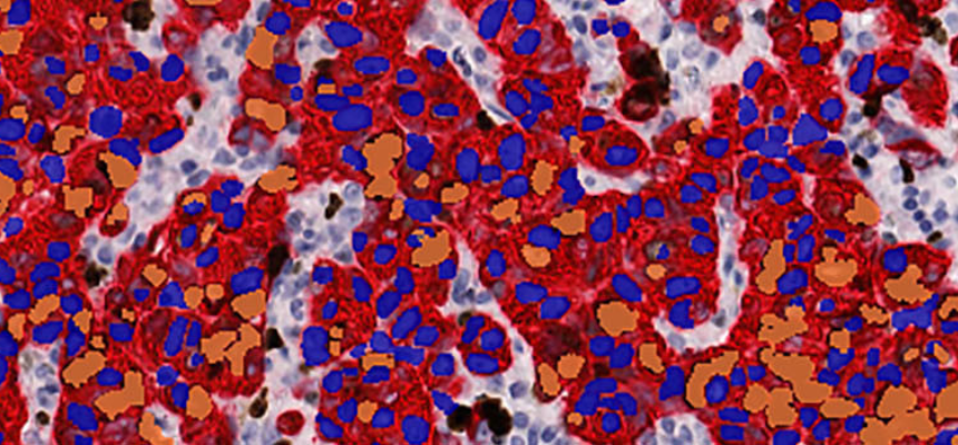
Visiopharm Invasive Tumor Detection (VDS)
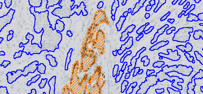
Microscopic view of breast tissue with invasive tumor area marked with orange hatching and blue contours.

RUO

IHC

Breast

Visiopharm

LEARN MORE

Visiopharm Iron Hematoxylin, Adipose Tissue
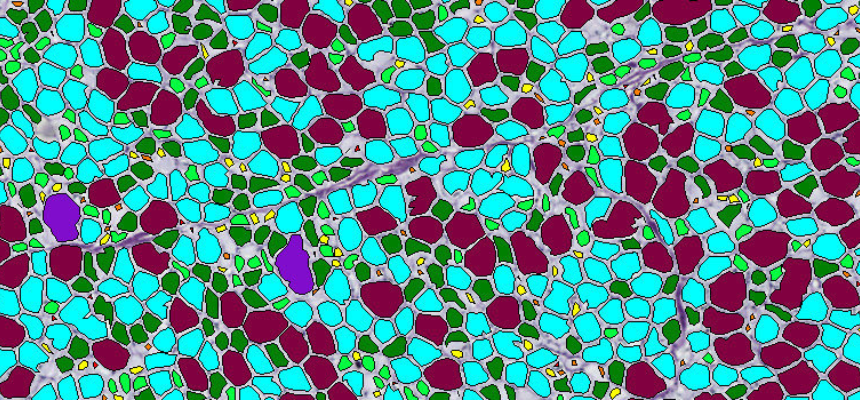
Microscopic image with segmented cells in turquoise, purple, green, and yellow outlined in black.

RUO

Other

Various

Visiopharm

LEARN MORE

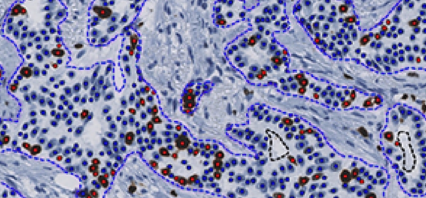
Visiopharm Ki-67

Microscopic tissue sample with segmented cell nuclei marked by red dots and outlined in blue.

CE-IVD

IHC

Breast

Visiopharm

LEARN MORE

Visiopharm Ki-67 + CK5 VDS, Neck Cancer
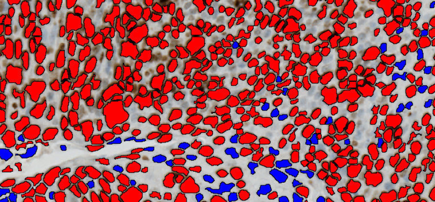
Microscopic image showing clustered cells highlighted in red and scattered cells in blue with black outlines.

RUO

IHC

Head & Neck

Visiopharm

LEARN MORE

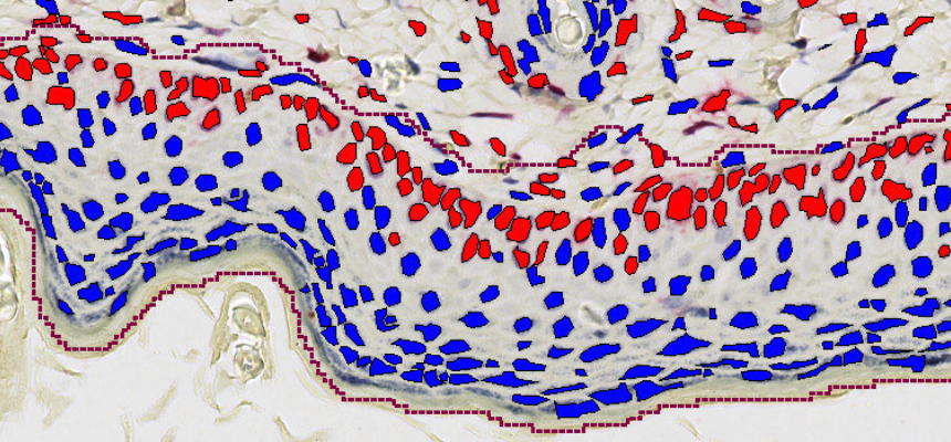
Visiopharm Ki-67, Epidermal Hyperplasia

Microscopic skin tissue section with cell nuclei marked in red and blue within dotted purple boundaries.

RUO

IHC

Skin

Visiopharm

LEARN MORE

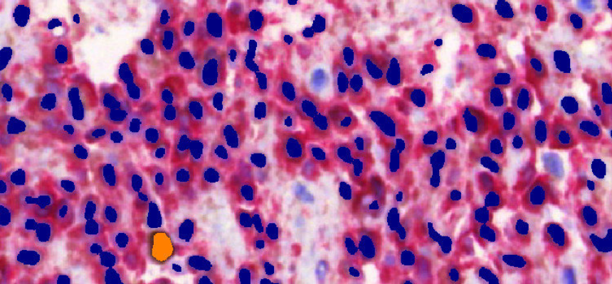
Visiopharm Ki-67+MART1, Melanoma

Microscopic image showing densely packed dark blue and red stained cells with one orange cell.

RUO

IHC

Skin

Visiopharm

LEARN MORE

Visiopharm Ki-67, Neuroendocrine Neoplasm
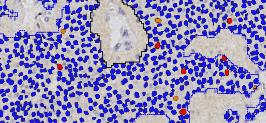
Microscopic tissue image with blue, red, and orange segmented cell nuclei outlined in black and blue.

RUO

IHC

Neuroendocrine

Visiopharm

LEARN MORE

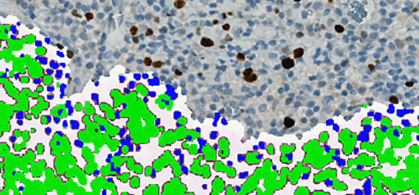
Visiopharm Ki-67+p16, Cervical Cancer

Microscopic image of cervical tissue with highlighted green and blue cells against brown-stained nuclei.

RUO

IHC

Cervical

Visiopharm

LEARN MORE

Visiopharm Ki-67+PanCK, Breast Cancer

Microscopic image of breast cancer tissue with red, blue, and brown stained cells visible.

RUO

IHC

Breast

Visiopharm

LEARN MORE

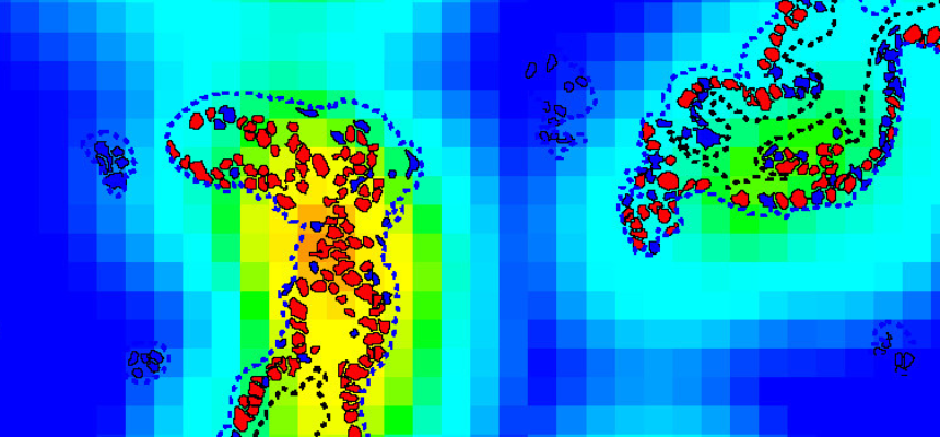
Visiopharm Ki-67, Pancreatic Cancer

Heat map with blue, green, yellow gradient and red markers highlighting clustered areas inside dotted boundaries.

RUO

IHC

Pancreas

Visiopharm

LEARN MORE

Visiopharm mCherry+GFP, Axon Density
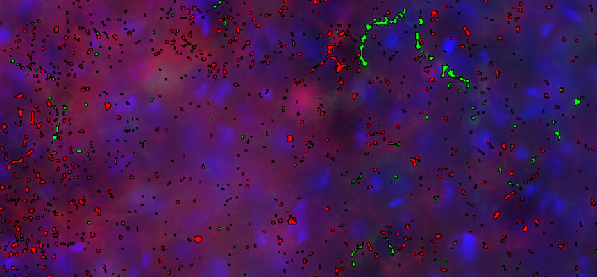
Microscopic image showing scattered red and green highlighted cells or structures on a blue background.

RUO

Other

Brain & Nervous System

Visiopharm

LEARN MORE

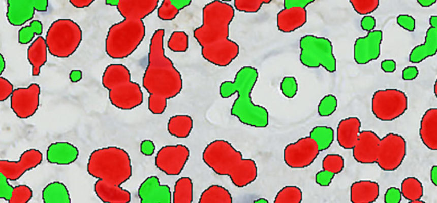
Visiopharm MCM7, Bladder Cancer

Microscopic view showing scattered red and green irregular shapes on a pale background.

RUO

IHC

Bladder

Visiopharm

LEARN MORE
No results found.
There are no results with this criteria. Try changing your search.

AI in your workflow

Image analysis AI applications work seamlessly with the Concentriq review experience.

Pathologists and scientists can see results update as they are available,  and use granular overlay tools to inspect and confirm results with confidence. They stay focused and in control without slowing down.
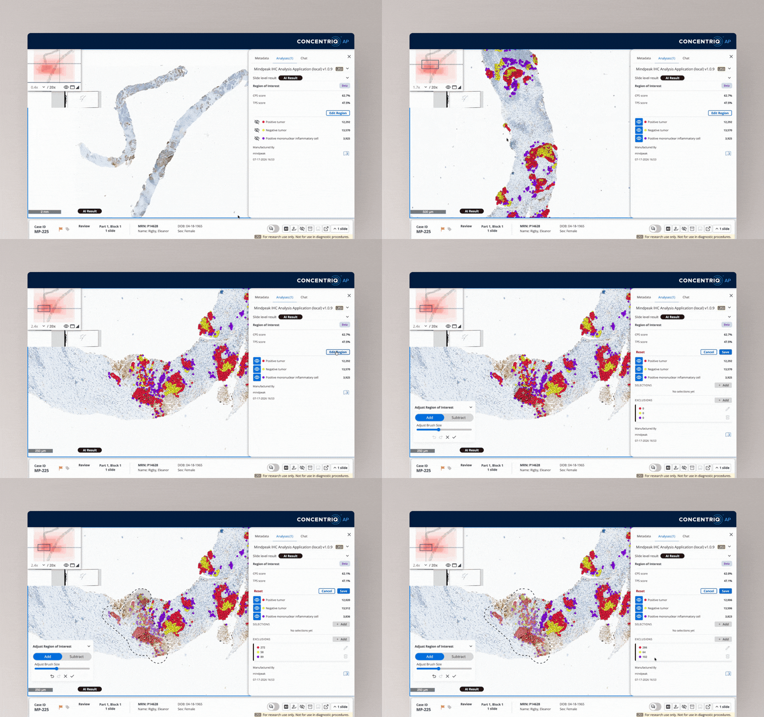
Digital histology slide showing stained tissue analysis with tumor and inflammatory cell detection results.

Latest Resources