The Precision Medicine AI Portfolio
Browse the largest curated AI ecosystem for pathology. All applications are available through Proscia and are interoperable with Concentriq. Reach out to see a demo, and talk about AI strategy for your lab.


Filters
Tag
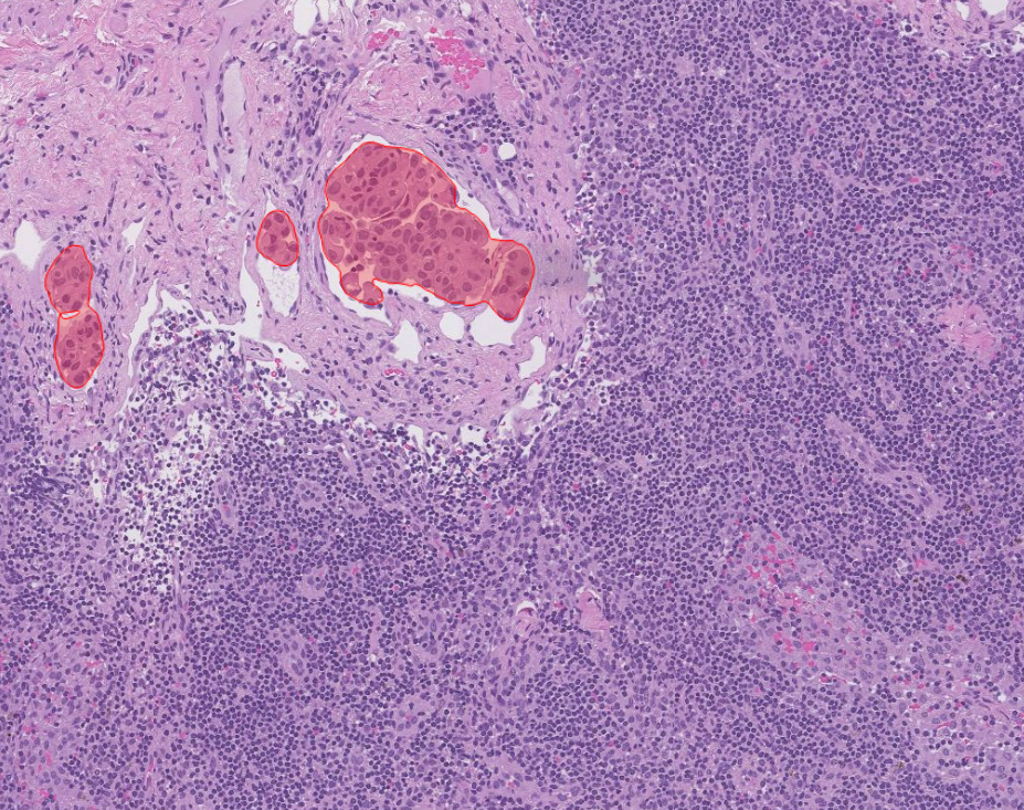
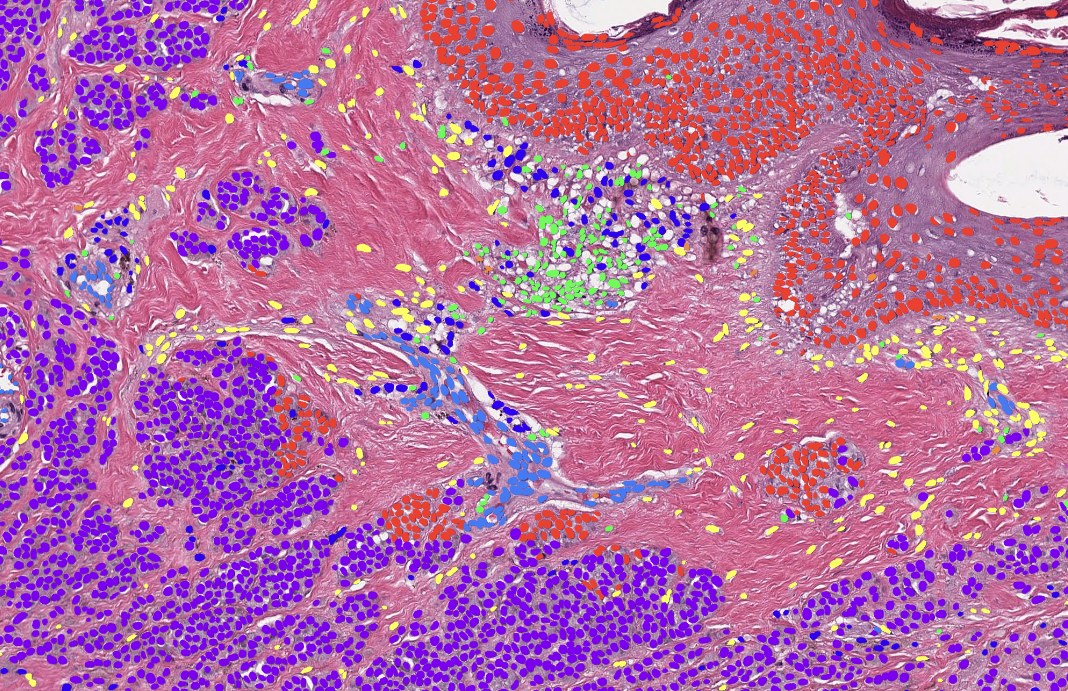
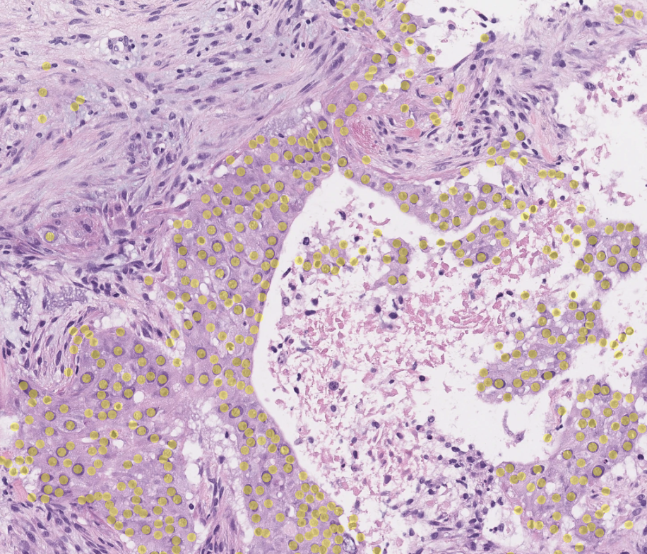
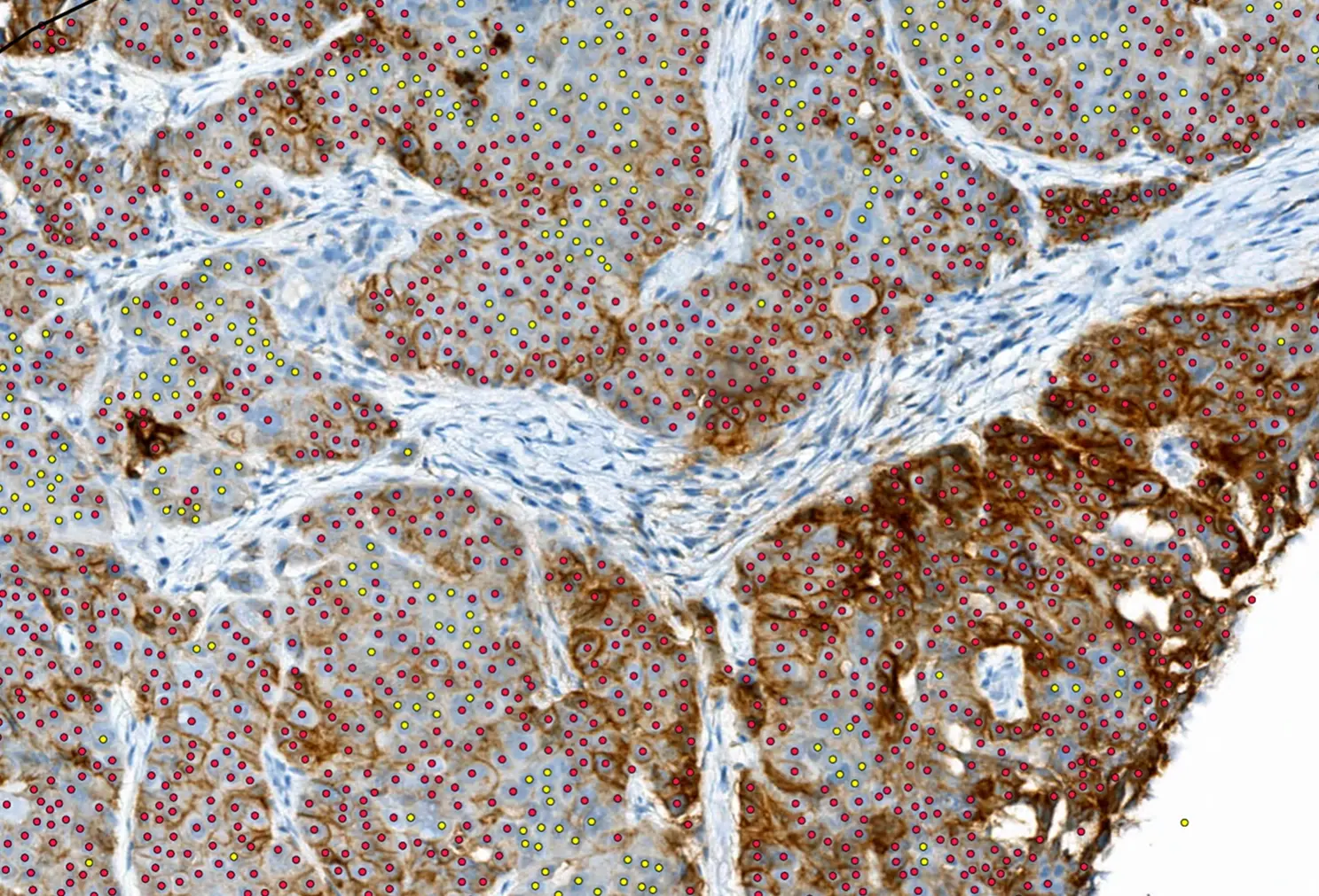
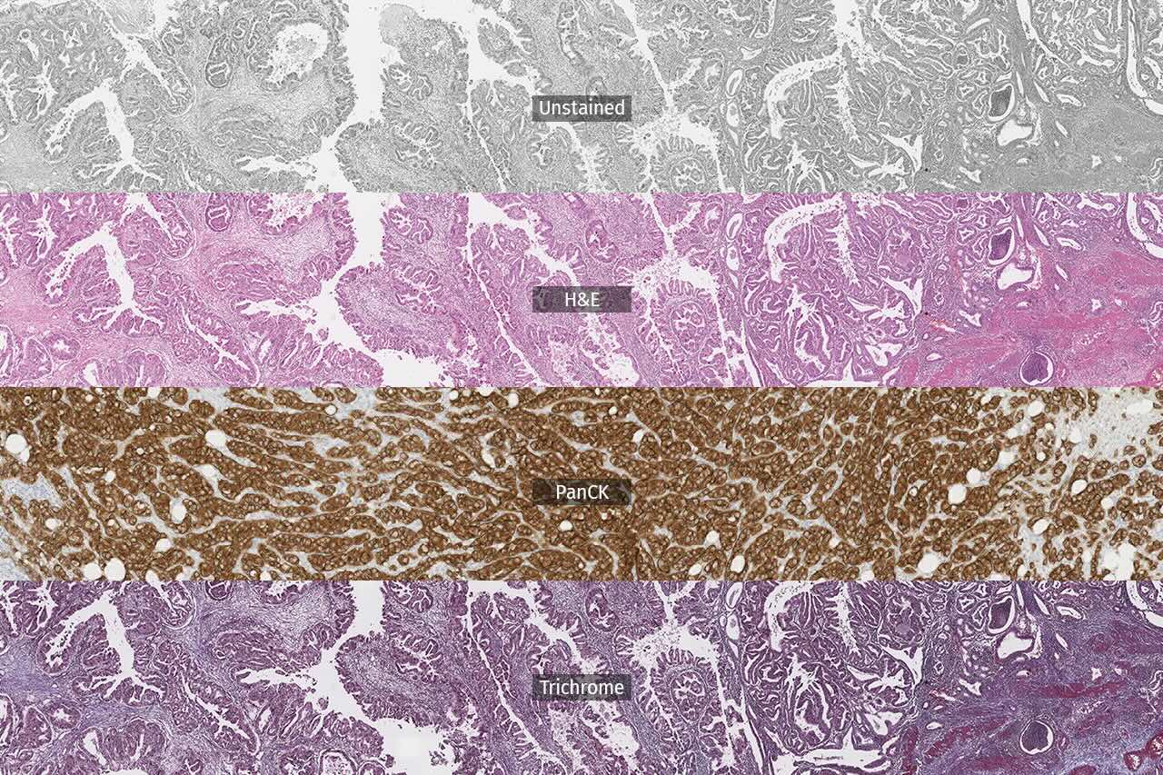
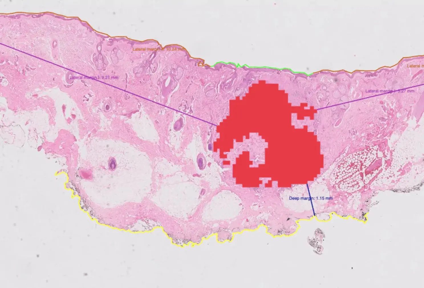
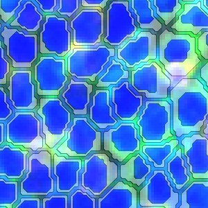
Visiopharm Cell Detection, AI (Fluorescence)
LABEL
RUO
STAIN
Other
TISSUE TYPE
Various
VENDOR
Visiopharm
No results found.
There are no results with this criteria. Try changing your search.
AI in your workflow
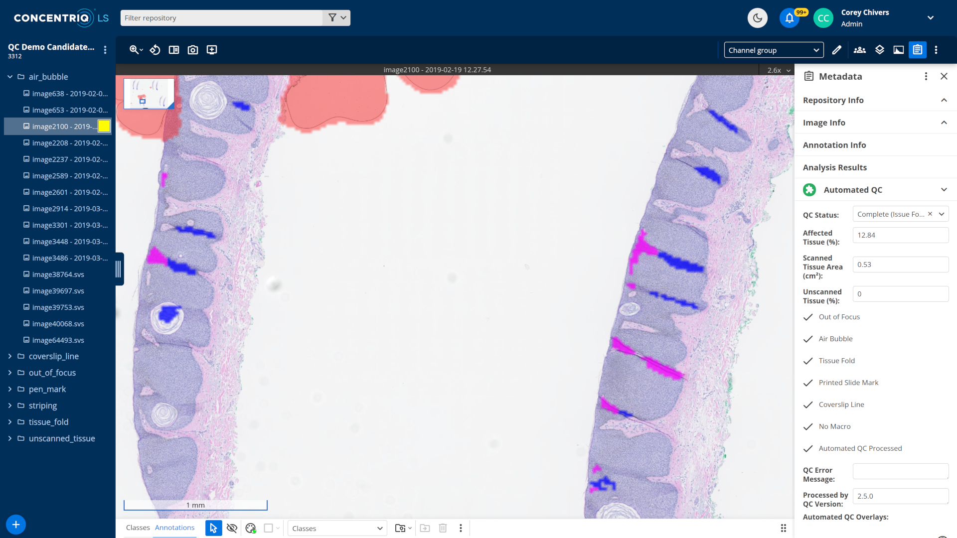
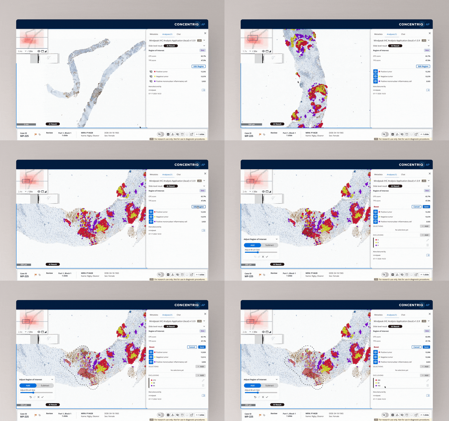
Image analysis AI applications work seamlessly with the Concentriq review experience.
Pathologists and scientists can see results update as they are available, and use granular overlay tools to inspect and confirm results with confidence. They stay focused and in control without slowing down.
“We wanted to be AI agnostic and be able to seamlessly play in that space with one viewer, and we are doing that.”



VP/Other Executive
KLAS Research, June 2024